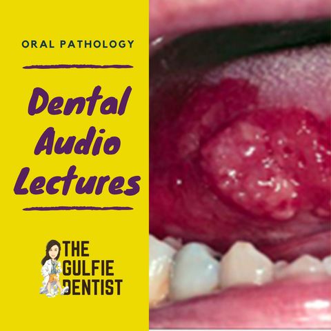
2. Development disorders

Download and listen anywhere
Download your favorite episodes and enjoy them, wherever you are! Sign up or log in now to access offline listening.
Description
DEVELOPMENTAL CONDITIONS Developmental conditions are soft tissue or hard tissue defects that occur during the development of the individual, either before or after birth. LIP PITS - Depressions or concavities...
show moreDevelopmental conditions are soft tissue or hard tissue defects that occur during the development of the individual, either before or after birth.
LIP PITS
- Depressions or concavities seen on lip
- Seen with Van Der Wood syndrome along with cleft lip
FORDYCES GRANULES
- Ectopic sebaceous glands
- On buccal mucosa
- Usually seen as Bilaterally symmetrical
LEUKOEDEMA
- White or whitish grey edematous (fluid) lesion of buccal mucosa
- It dissssipiates when cheek is stretched
ANGIOMAS
- ANGIO - VESSELA OMA - TUMOR
- TUMORS composed of blood vessels or lymph vesseLs
- A salivary gland tumor that mestasises to bone
CENTRAL HEMANGIOMA – IV
- Commonly in upper lip
- There is congenital focal proliferation of capillaries
- Absolute contradiction – extraction of a tooth**
- Multilocular radiolucency lesion
- Associated syndrome – Struge Weber Syndrome
- Port wein steins + calcification of duramatter
- Strawberry appearance of skin – capillary hemangioma
Other variants
- Strawberry appearance of gingiva – warners granulomatosum
- Strawberry appearance of tongue (white coated tongue with red inflamed fungiform papilla)– scarlet fever (bacterial infection)
- Raspberry appearance of plate – papillary hyperplasia(denture)
LYMPHANGIOMA
CONGENITAL FOCAL PROLIFERATION OF LYMPH VESSELS
-Oral lymphangiomas are very rare
- appear as purple spots on tongue
DEVELOPMENTAL SOFT TISSUE CYST - DERMOID CYST
- Mass in the midline of the body – intraoral or extra oral
- Intraorally – floor of the mouth if above mylohyoid
- Mass will be seen in the upper neck if it forms below the mylohyoid
- Contains hair, sebaceous glands etc – doughy consistency
BRANCHIAL CYST
- Lateral neck cyst
- Epithelial cyst within lymph node of neck
PSEUDOCYSTS OF JAW
STAFNE / STATIC BONE CYST
- Radiolucency in the post mandible below the mandibular canal
- Its is not a cyst- just a picture caused due to lingual concavity of the jaw, ie. An invagination in the lingual surface of the jaw - just variation of normal anatomy.
NASOPALATINE / MEDIAN PALATINE / Incisive canal cyst CYST
- Seen b/w roots of Central incisors
- R/F - Divergence of roots
- The nasopalatine cyst appears as a well-defined, round radiolucency in the midline of the anterior maxilla
- Sometimes it appears to be ‘heart-shaped’ because of superimposition of the anterior nasal spine.
- Radiological assessment should include examination of the lamina dura of the central incisors (to exclude a radicular cyst) and assessment of size (the nasopalatine foramen may reach a width of as much as 10 mm).
- Qn. Pt. came to the clinic complaining from pain related to swelling on maxillary central incisor area with vital (under percussion) - nasopalatine cyst
- Qn. 40-60y, Male, in maxilla in the midline between the roots of upper central incisors which are vital.
- Intra-osseous lesion is well circumscribed rounded or Heart-shape RL. area (due to superimposition of nasal spine)
GLOBULOMAXILLARY CYST
- Seen between Lateral incisor and Canine
- Inverted pear shape
- Variant of OKC or lateral periodontal cyst
- Vital tooth bilateral
SOLITARY BONE CYST – Haemorrhagic /Simple / Traumatic bone cyst
- A pseudocyst- ie. No epithelial lining
- Kids doing sports- injury
- R/F – scalloped border around teeth***
- Treatment is curettage and closure
Information
| Author | The Gulfie Dentist |
| Organization | The Gulfie Dentist |
| Website | - |
| Tags |
-
|
Copyright 2024 - Spreaker Inc. an iHeartMedia Company

Comments